



Giá đỡ điện thoại cho kính hiển vi (Microscope Phone Holder)
 Byleizerdesign_249154Likes62Downloads801
Build another version of this model.
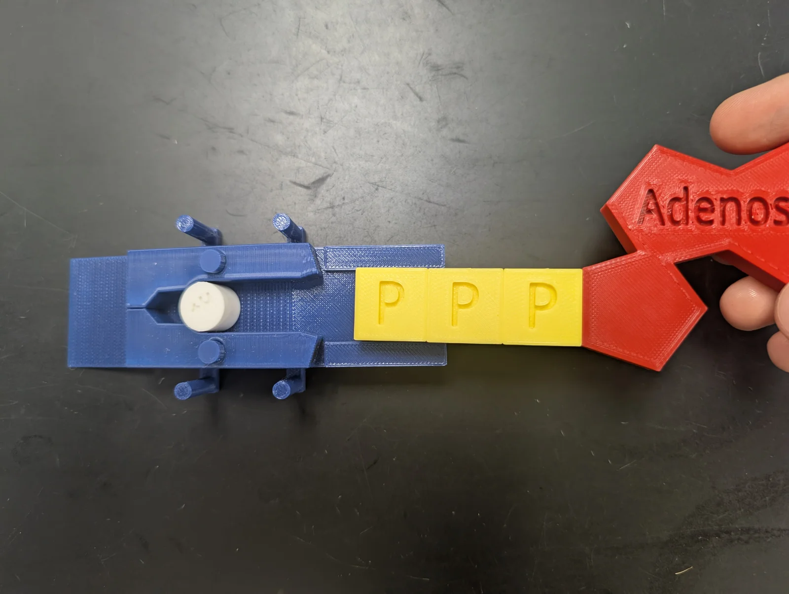
This model is intended to be used with a Magnetic ATP Model.
The arms should be printed in a vertical orientation to minimize supports. The central hole in each arm may require some cleaning out of all support material to ensure it can pivot on the central posts of the protein base. Each arm needs a 4x2mm disc magnet embedded it the enlarged end, and each arm should have its magnet oriented opposite to the magnet of the other arm. This creates enough attraction that the two ends will stick together at rest on the protein base. The narrow ends of each arm should be embedded with a 7x3mm disc magnet, oriented in the opposite polarity to the left-hand side of the phosphate groups produced using the Magnetic ATP Model.
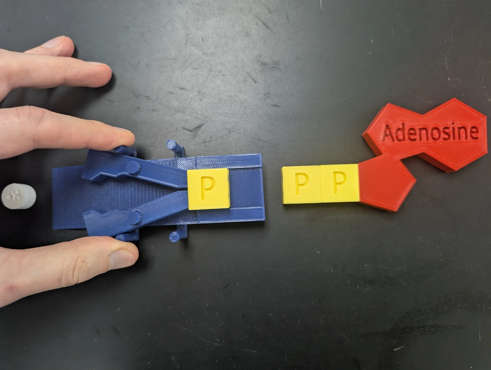
One H+ ion should be loaded into the protein pump at a time. If done correctly, when a phosphate group (like on an ATP molecule) is moved through the guiding lane on the protein base towards the arms, the arms will snap to the phosphate group. This allows the H+ ion to move through the pump. The change in shape of the protein pump reflects phosphorylation, the change in shape of a protein when a phosphate group is attached.
After the H+ ion has passed through, the phosphate group can be removed and the protein arms will return to their original position. This reflects dephosphorylation, the change in shape of a protein when a phosphate group is removed - and typically returns to its original shape.
Byleizerdesign_249154Likes62Downloads801

 ByBoonby_1059917Likes659Downloads1.2K

 ByFabianVeileLikes414Downloads1.5K

 BygillisLikes121Downloads2.1K

 ByMarciLikes87Downloads295

 Bystrifu_4061046Likes151Downloads1.3K

 ByStobbartLikes15Downloads53


 ByzoltanfLikes73Downloads276


 ByKavelry3D_844921Likes5Downloads36

 ByKiwis Cant Fly Cant FlyLikes0Downloads6